Registered with the Registrar of Newspapers for India under R.N.I 53640/91
Vol. XXXI No. 14, November 1-15, 2021
A papier‑maché human anatomical model in the Madras Medical Establishment in the 1830s
by Ramya Raman and Anantanarayanan Raman
An untitled 2-page note by John Carnac Morris,1 signed ‘ED.’ at the end, sleeved under ‘Scientific Intelligence’ in the inaugural issue of the Madras Journal of Literature & Science [MJLS] (Morris, 1834) refers to a human-anatomical model in Madras (Fig. 1). Made by a French physician, Louis Auzoux, this model was noticed in the MJLS because of its ‘interactive’ endowment and near-human size. The model was unique, because it could be dismantled to view internal body parts and re-fitted snugly to its normal human appearance. Thus it was amenable for learners of medicine in an interactive manner.
The Madras Medical School (M.M.S.) formally commenced in February 1835, shortly after Morris’s above-referred notice. The General Hospital in Madras (M.G.H.) was operating for army personnel from 1772, which started serving the general public in the 1840s. Before the establishment of M.M.S., the M.G.H. trained Europeans, Eurasians (Anglo-Indians), and Indians in western methods of diagnosis and treatment and pharmacopoeiae for people to serve under the Subordinate Medical Service (S.M.S.) of Madras in eighteenth and nineteenth centuries. The Subordinate Medical Servants trained as either Apothecaries or Dressers were employed in dispensaries in rural and remote villages and hospitals in major towns of the then Madras Presidency as assistants to British-qualified doctors. William Mortimer superintended the M.G.H. from 1827, who additionally held charge of M.M.S., when it was commissioned newly in February 1835. The present article refers to Morris’s report on the interactive model brought to Madras in 1832, highly likely for use in training the S.M.S. candidates.
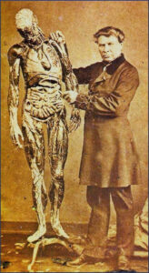
Fig. 1: Louis Auzoux and life-size human model, 1845 [Museo Nacional de
Medicina, Quito, Ecuador]. Possibly a similar model arrived in Madras in 1832.
John Morris’s note
Morris describes the model as an ‘anatomical figure’ and an ‘exact model of the human body, with the skin taken off’ constructed by ‘Chevalier Auzoux’ (Morris, 1834). He says: “It (the model) represents, with the greatest minuteness, and critical precision, all the details of the human structure. Each muscle and organ can at pleasure be removed and examined, with the same results that are attainable by the process of dissection. Indeed whether as regards its striking resemblance of nature, or the anatomical correctness of its construction, it may be considered one of the most extraordinary combinations of art and science, that have appeared in the present day.”
This model was brought to Madras from England by one Mr. Knox. An identical model was procured – may be a little earlier, date not available – for medical training in Calcutta. Morris also says that Knox published a pamphlet on the relevance and use of such models in medical schools. That pamphlet, unfortunately, is not traceable, although its bibliographic details were, shown below:
Knox, G., 1834. Description of an artificial anatomical figure, constructed by Chevalier Auzoux, M.D. exhibited in 1832 before the King in London, Church Mission Press, Madras.
While searching for Knox’s pamphlet referred to by Morris (1834), we chanced upon The Anatomist’s Instructor and Museum Companion (1836) by another Knox – Frederick John Knox – who qualified for the Licentiate of the Royal College of Surgeons of Edinburgh in 1831, immigrated to New Zealand in 1840, and practiced there as a surgeon. The Frederick Knox volume published in 1836 also refers to Auzoux’s anatomical models.
George Knox in Morris’s note
The Knox referred in Morris’s note was George Knox, a surgeon who worked with the Madras Medical Establishment in the 1830s. George Knox was an Indian Medical Servant. He was a Fellow of the Royal College of Surgeons of London from 1816 (The Royal College of Physicians of London, 1845). Except a communication published in the Royal Asiatic Society of Great Britain and Ireland (1831), no other documented work of George Knox exists.
Louis Auzoux in Morris’s note
Louis Thomas Jerome Auzoux (1787–1880) was a qualified French medical doctor, who studied with Guillaume Dupuy and Rene-Alexis Baffos at the Paris Medical School in 1818 and 1820, respectively. Human anatomy attracted Auzoux. Serendipitously, Auzoux, while studying in Paris, ran into an elderly lady selling papier-maché dolls in Paris streets. Papier-mâché, as a material, impressed Aujoux as suitable for his proposed human-anatomical models, since, he considered that it would be soft and resistant, and not stick to the cast (Olry, 2000). The French Government decorated Auzoux with the title ‘Chevalier’.
Auzoux pioneered in creating papier-mâché models casting them in lead matrix under a heavy wood coating. The courses of nerves and blood vessels were articulated by gluing hemp fibres, subsequently hand painted (Olry, 2000). Auzoux called these creations as clastics deriving from klastos (Greek) meaning ‘in pieces’. At l’Académie Nationale de Médecine (l’ANM, the National Academy of Medicine), Paris, Auzoux exhibited his first model of a human-lower limb in 1822. l’ANM constituted a committee comprising leading medical professionals of Paris, viz., Nicholas-Philibert Adelon, Anthony Dubois, Francois Ribes, Hippolyte Cloquet, Jean Cruveilhier, Gilbert Breschet, and Rene-Alexis Baffos to verify Auzoux’s creations and explanations in 1825–1830. The Adelon committee submitted a formal report in 1831 (Adelon et al., 1840) to l’ANM, zealously endorsing Auzoux’s creations:
Your favourable opinion, gentlemen, has been confirmed by the extraordinary anxiety evinced by the public institutions of almost all civilized countries to obtain this wonderful preparation. Your elogiums and the avidity with which these specimens have been sought after by Foreigners, have given a new impulse to the zeal of our colle(a)gue (sic. Auzoux). We transcribe, with great pleasure, what was said in 1823 by the Medical Society of Emulation, “We cheerfully accord to M. Anzoux the thanks due to his zeal in the cause of science.” his patience, his ingenious essays, and the brilliant results accomplished by his perseverance and profound knowledge of anatomy.
The Adelon committee examined a human model measuring 5 feet 6 inches (c. 170 cm) made by Auzoux. According to them, this model delineated every minute detail of the structure of human body. The following passage from their report reinforces their satisfaction on Auzoux’s submission:
The representation of the Heart is exceedingly happy; by means of a section made in the inter-auricular and inter-ventricular partition, this organ is divided into two halves: upon each half are two cavities, which may be opened so as to bring into view the valves — all these parts re-unite so exactly, that the traces of division can scarcely be recognized — and in the entire they exhibit a heart of natural size, whence are seen the vessels, which originate from this organ or returned to it. All these vessels being traced from their origin to their termination it is easy to study their different branches, their numerous anastomoses, and their relation with the different organs.
1 John Morris, a Madras Civil Servant, edited Madras Journal of Literature & Science journal, from 1834 to 1836, who was succeeded by Robert Cole. Morris contributed to the understanding of Telugu language and literature. He was admitted to the Fellowship of Royal Society in 1831. He was afflicted by paraplegia at a relatively young age. He worked in Madras confined to a chair in Madras Government Secretariat.
Anantanarayanan Raman: Charles Sturt University, PO Box 883, Orange, NSW 2800, Australia; CSIRO (Health & Biosecurity), Floreat Park, WA 6014. Australia.araman@csu.edu.au; anant@raman.id.au
Ramya Raman: School of Medicine, University of Notre Dame, Fremantle, WA 6160, Australia.
ramya.raman@nd.edu.au
(To be continued next fortnight)

Fascinating. Reminds me of the complicated three dimensional books made in Europe in 17th century to teach medicine. As explained by Jay Walker – https://youtu.be/P-SRD4qal7Q
Pingback: A papier‑maché human anatomical model in the Madras Medical Establishment – II « Madras Musings | We Care for Madras that is Chennai